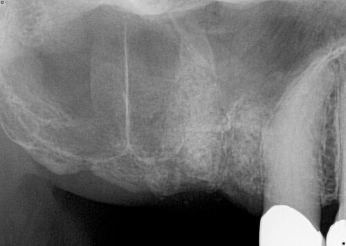
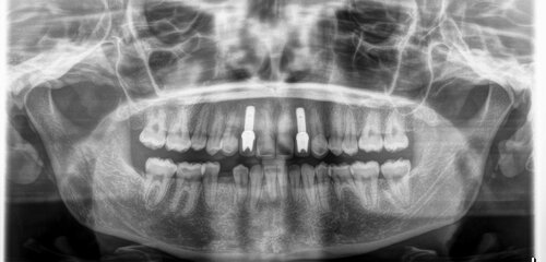
The upper back molars were missing and the sinus was too low to proceed with dental implants. There was not enough bone present to place the implant.
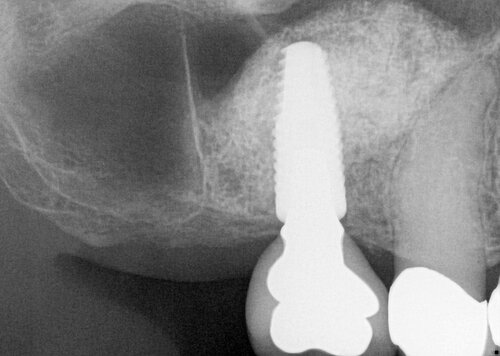
After
We performed a sinus augmentation procedure to “lift” the sinus and place bone into the site so that a dental implant could be anchored properly, restoring molar function.
Before
This patient was congenitally missing these front teeth, which never developed.
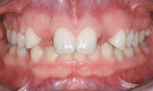
During
We placed two narrow dental implants into the bone to replace the missing tooth roots.
After
The patient received a wonderful aesthetic and functional result with porcelain crowns over the implant fixtures.
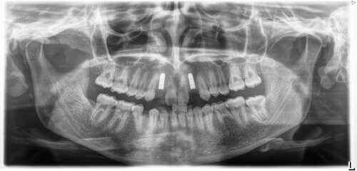
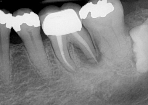
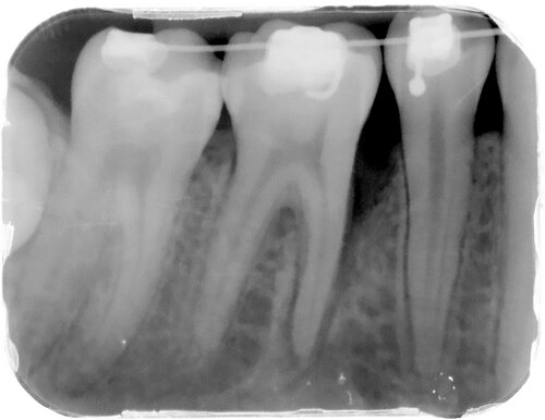
Before
The root of this molar was fractured, and the tooth had to be removed to prevent further bone loss and infection.
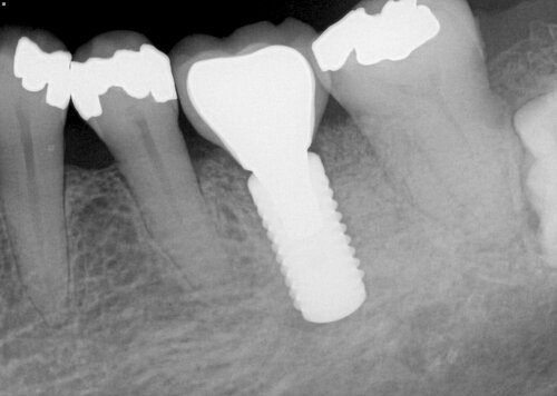
After
A dental implants placed into the molar site was able to restore the original function to the area. This also helped to avoid tipping and migration of the adjacent teeth. Having an implant is often preferable to a bridge because it avoids having to cut the adjacent teeth down for crowns, and implants are easier to clean around. In addition, implants do not get cavities.
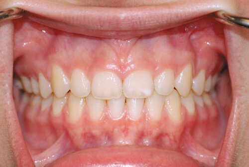
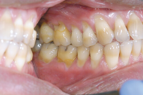
Before
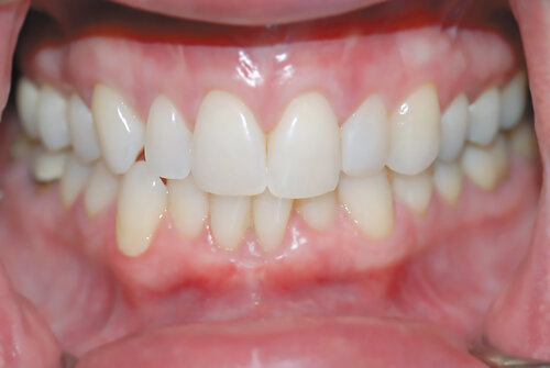
The tooth length of the upper front teeth appears too short, making the teeth appear square shaped and showing too much gum tissue when the patient smiles.
After
We performed a simple gum lift procedure called crown lengthening to improve the shape of the teeth. The result makes them longer and more triangular, which is an aesthetic improvement.
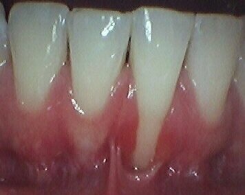
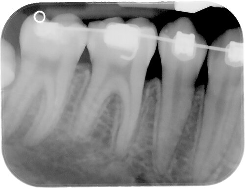
Before
This patient shows severe recession (exposed root surfaces) of the lower front teeth. They lacked the band of thick gum tissue that should be present and instead had only mucosa. This mucosal tissue is thin and mobile (like the inside of your lip), and does not create a protective seal against the tooth.
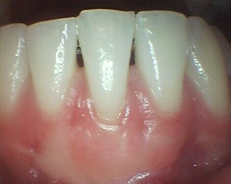
After
We performed a gum graft utilizing tissue transplanted from the roof of the mouth. The patient achieved a thick, stable long-term result that created a firm protective seal against the tooth to prevent further breakdown.
Before
Note the exposed root surfaces (recession) of the upper teeth.
After
We performed a simple gum grafting procedure to reposition the gum tissue and cover the roots. This helps decrease the chances of root cavities and sensitivity while providing significant aesthetic improvement.
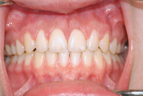
Before
Every tooth exhibited exposed roots, called recession. Exposed root surfaces are softer than enamel. That makes the teeth more prone to cavities, abrasion, and sensitivity.
After
We used donor gum tissue to treat all of these teeth at once in a procedure called gum grafting. The roots were covered, making them more resistant to long-term problems while improving aesthetics.
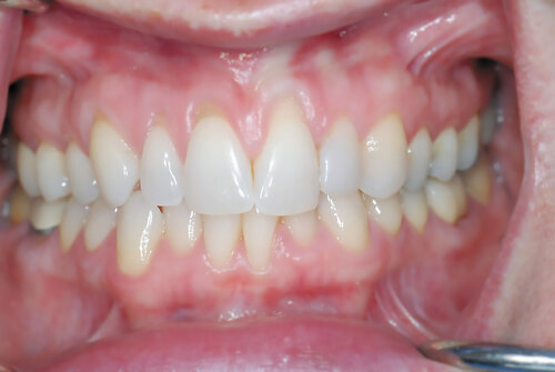
Before
The patient developed severe bone loss while undergoing orthodontic treatment (braces). This type of defect is high risk for tooth loss if left untreated.
After
Minimally invasive laser surgery disinfected the site and stimulated bone growth. After one year the patient enjoyed complete resolution of the defect.